SpectraPro HRS Imaging Spectrographs and Scanning Monochromators

Princeton Instruments SpectraPro HRS builds on the 25 year legacy of reliability and precision of the original SpectraPro series of imaging spectrographs and scanning monochromators. Available in 300mm, 500mm and 750mm focal lengths with wide variety of input and output configurations.


Overview

SpectraPro HRS builds on the 25 year legacy of reliability and precision of the original SpectraPro series of imaging spectrographs and scanning monochromators. Teledyne Princeton Instruments has created new feature-packed, higher resolution models ready for any application.

Powered by improved optical design, innovative software and exclusive technologies, SpectraPro HRS are unmatched in performance and versatility.

There are 3 focal lengths, 300mm, 500mm and 750mm. Each is available in multiple configurations with dual exit ports, single or dual entrance slits and dual exit slits. All are compatible with the new range of application CUBES and a wide range of accessories.

ResXtreme Spectral Deconvolution

  • Improves spectral resolution by up to 60%
  • Improves peak intensity of spectral lines by up to 60%
  • Conservation of Energy: Maintains total signal under the peak
  • Improves signal-to-noise ratio
  • Preserves system throughput
  • included with all SpectraPro HRS spectrographs that are purchased with Princeton Instruments LightField software!

AccuDrive Triple Grating Turret

  • Dramatically improved wavelength accuracy and repeatability
  • Outperforms previous scan systems – yields significant improvements in accuracy and reproducibility
  • Automatically identifies the turret and gratings installed on startup
  • Performs several optical alignment routines to insure accurate initialization

Powered by LightField

  • Optional: LightField® (for Windows® 7/8, 64-bit)
  • LightField offers intuitive, cutting-edge user interface, IntelliCal, hardware time stamping, and more.

Specifications

Specifications HRS-300 HRS-500 HRS-750
Focal length 300 mm 500 mm 750 mm
Aperture ratio f/3.9 f/6.5 f/9.7
Spectral resolution with PMT* 0.09 nm with 10 μm wide slits 0.05 nm with 10 μm wide slits 0.03 nm with 10 μm wide slits
CCD spectral resolution** 0.10 nm 0.07 nm 0.05 nm
CCD spectral resolution with ResXtreme 0.07 nm or better (typ.) 0.05 nm or better (typ.) 0.03 nm or better (typ.)
Reciprocal linear dispersion 2.38 nm/mm 1.52 nm/mm 1.03 nm/mm
Wavelength coverage across
26.8 mm wide CCD
64 nm (nominal) 41 nm (nominal) 27 nm (nominal)
Focal plane size 14 mm x 30 mm (both exit ports)
Scan range 0 to 1500 nm
Drive step size 0.002 nm/step
Wavelength accuracy +/-0.2 nm (up to 0.02 nm with IntelliCal wavelength calibration) +/-0.01 nm
Wavelength reproducibility +/-0.05 nm (both scan directions, typ.)
Turret Triple-grating interchangeable CTS-Turrets self-align to system when installed
Grating change repeatability 0.02 nm (typ.)
Grating size 68 mm x 68 mm standard; 68 mm x 84 mm optional
Number of turrets allowed Accepts as many as 3 turrets, each with 3 gratings
Astigmatism correction Toroidal mirror reduces astigmatism to permit multichannel spectroscopy
Computer interface USB 2.0
Certification CE tested to the following standards: EN 55022:2010/AC:2011, EN 61000-3-2:2014, EN 61000-3-3:2013, EN 61000-6-3:2007/A1:2011, EN 61326-1:2013
Dimensions
Length 15.71” (399 mm) 22.13” (562 mm) 31.75” (806 mm)
Width 11.00” (279 mm) 11.84” (301 mm) 12.50” (318 mm)
Height 8.74” (222 mm) 8.74” (222 mm) 9.16” (233 mm)
Weight 43 lbs (19.5 kg) 50 lbs (22.7 kg) 75 lbs (34.0 Kg)
Optical axis height 6.155” ± 0.435” (156 mm +/- 11 mm), adjustable
Note: Unless otherwise noted, specifications with a 1200 g/mm grating @ 435.8 nm Specifications are subject to change.
* PMT resolution measured with a 1200 g/mm grating @ 435.8 nm, 10 μm slit width, and 4 mm slit height.
** CCD resolution measured at the focal plane center with 10 µm slit and a Princeton Instruments PIXIS:400F with 20 µm pixels @ 546 nm.
For astigmatism-free performance, please see our IsoPlane-320 and 160 spectrographs.

Applications

Tip-Enhanced Raman Spectroscopy
TERS – Tip-Enhanced Raman spectroscopy

Laser-Induced Breakdown Spectroscopy
LIBS is considered one of the most convenient and efficient analytical techniques for trace elemental analysis in gases, solids, and liquids. LIBS spectra obtained by the Mars Curiosity Rover have confirmed that our sister planet could have harbored life

Fluorescence, Phosphorescence, and Photoluminescence Spectroscopy
Fluorescence, phosphorescence and photoluminescence occur when a sample is excited by absorbing photons and then emits them with a decay time that is characteristic of the sample environment.

Astronomical Imaging
Astronomical imaging can be broadly divided into two categories: (1) steady-state imaging, in which long exposures are required to capture ultra-low-light-level objects, and (2) time-resolved photometry, in which integration times range from milliseconds to a few seconds.

General Raman
The most common application of Raman spectroscopy involves the vibrational energy levels of a molecule. Incident laser light in the UV, visible or NIR, is scattered from molecular vibrational modes.

Surface-Enhanced Raman Spectroscopy
SERS – Surface-enhanced Raman spectroscopy

Coherent Anti-Stokes Raman Spectroscopy
Coherent Anti-Stokes Raman spectroscopy (CARS) a type of non-linear Raman spectroscopy. Instead of the traditional single laser, two very strong collinear lasers irradiate a sample.

Combustion
Combustion researchers rely on laser-based optical diagnostic techniques as essential tools in understanding and improving the combustion process.

Singlet Oxygen Imaging
Singlet oxygen, the first excited state of molecular oxygen, is a highly reactive species that plays an important role in a wide range of biological processes, including cell signaling, immune response, macromolecule degradation, and elimination of neoplastic tissue during photodynamic therapy.

Plasma Emission Spectroscopy
The different types of plasma emission spectroscopy can be categorized by how the plasma is generated. Spectra of nuclear fusion plasmas are used to ascertain the chemical species present and other properties.

Resonance Raman Spectroscopy
Instead of fluorescence, some types of colored molecules produce strong Raman scattering at certain conditions. This effect was called Resonance Raman.

Stimulated Raman Scattering
Stimulated Raman scattering takes place when an excess of Stokes photons that were previously generated by normal Raman scattering are present or are deliberately added to the excitation beam.

Resources

Datasheets:

Datasheet

Videos:

Fundamentals of Spectroscopy

Technical Notes:

IntelliCal-Automated wavelength and intensity calibration routines significantly improve accuracy of recorded spectra
Automated wavelength and intensity calibration routines significantly improve accuracy of recorded spectra.

Fully automated wavelength calibration method optimizes data accuracy
Patent-pending IntelliCal® calibration technology from Princeton Instruments enables fast, reliable wavelength calibration with minimal user input.

Better Imaging with a Schmidt-Czerny-Turner Spectrograph
The IsoPlane spectrograph has a unique optical design that completely eliminates field astigmatism at all wavelengths and at all points across the focal plane, and reduces coma to negligible levels. This means the IsoPlane gives sharp and spatially well resolved images across the entire CCD sensor.

Improved Spectra with a Schmidt-Czerny-Turner Spectrograph
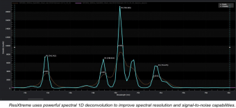
The data in this paper have shown that by decreasing optical aberrations and increasing fluence, the IsoPlane gives spectra with better spectral resolution and SNR compared to Czerny-Turner spectrographs. Higher spectral resolution means peaks that are too close together to be resolved by a CT spectrograph can be clearly seen with the IsoPlane.

Application Notes:

Analysis of Perovskite Solar Cells via Spectral Luminescence in the 700 to 1000 nm Wavelength Range
03/23/2018  The rapid development trajectory of perovskite solar cells (PSCs) is attributable to a multitude of ongoing research efforts that target technological challenges associated primarily with device stability and process uniformity. The ability to detect efficiency-limiting defects, for example, is playing a particularly critical role in both PSC fabrication and performance improvements.

Low-Frequency Raman Spectra of Amino Acids Measured with an Astigmatism-Free Schmidt-Czerny-Turner Spectrograph: Discovery of a Second Fingerprint Region

Microscopy and Raman Imaging: Open-system Raman microscopy
Author: Cynthia Hanson and Elizabeth Vargis
05/05/2015  Publication: Laser Focus Wolrd
An IsoPlane 160 and a PIXIS 400 CCD camera are part of a cost effective Raman microscope solution developed at Utah State University. View the article in Laser Focus World.

Real-Time Imaging of Singlet Oxygen via Innovative Microspectroscopy Instrument
New Two-Dimensional InGaAs Detector Thermoelectrically Cooled to –85°C Facilitates Scientific Research

Scientific NIR-II/ SWIR Cameras Enable Femtosecond Frequency Comb Vernier Spectroscopy
New, Deeply Cooled InGaAs Cameras Provide Ultrahigh Sensitivity for Key Spectral Range

Aberration-Free Spectrographs and NIR-Sensitive InGaAs Cameras Facilitate the Development of Carbon Nanotube Optical Sensors for Early Disease Detection
Dr. Daniel Heller and his research group at Memorial Sloan Kettering Cancer center utilized the IsoPlane 320 spectrograph and NIRvana SWIR camera in their recent research.

Acquiring and processing Raman spectral data for the C2-D stretching vibration of 2 deuterated histidine
Because of histidine’s importance and unique functionality, we wanted to map out the probe group’s sensitivity to allow for its general use in protein related research.

Advanced CCD Cameras and Imaging Spectrographs Facilitate Acquisition of Novel Femtosecond Stimulated Raman Spectroscopy Data To Improve SERS Biosensors
Accurate characterization of surface-enhanced Raman spectroscopy (SERS) biosensors, fluorescent dye molecules that hold great promise for in vivo bioanalyte detection, can often be quite difficult as the overwhelming isoenergetic fluorescence signal typically makes it challenging to measure resonance Raman cross-sections for the molecules. To overcome this obstacle, researchers at the University of Minnesota in Minneapolis recently utilized etalonbased femtosecond stimulated Raman spectroscopy (FSRS), a technique designed to acquire a stimulated Raman signal without strong fluorescence or interference from signals resulting from other four-wave mixing pathway

Solar cell inspection via photoluminescence imaging in the NIR/SWIR
Scientific-grade, deep-cooled, large-format InGaAs FPA cameras such as the NIRvana from Princeton Instruments will enable researchers to observe photoluminescence emission at longer wavelengths and rapidly obtain more detailed information about defects within multicrystalline silicon solar cells.

Ultra-High-Speed, Time-Resolved Spontaneous Raman Scattering Spectroscopy in Combustion
The recent use of a new diagnostic apparatus to measure the dynamics of each individual molecular species, as opposed to simply acquire bulk information (e.g., pressure), points to the possibility of performing temperature and frequency analyses of species in combustion.

Low-frequency Raman Spectra of Carbon Nanotubes Measured with an Astigmatism-free Schmidt-Czerny-Turner Spectrograph
An IsoPlane has been interfaced to a low frequency Raman module that enables measurement of peaks with Raman shifts as low as 10 cm-1 using only a single stage spectrograph.

Accessories/ Related Products

Accessories

BLAZE® High-Speed Cameras for Spectroscopy

See CUBES

Request Info / Demo